
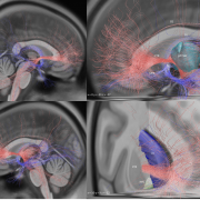

We’re really excited that the current cover of AoN was made with Lead-DBS. It shows pallidal and thalamic electrodes simultaneously implanted in a patient suffering from Tourette’s Syndrom and the picture is part of an electrophysiological Berlin-Hannover study spearheaded by Wolf-Julian Neumann and led by Andrea Kühn. It is the sixth coverart made with Lead-DBS that we know of – a gallery can be found here.


by our group &
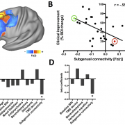
We just published a new study spearheaded by R. Ryan Darby that used the Lesion Network Mapping approach developed by Michael Fox to analyse lesion induced criminal behavior.
The study has been featured on multiple news outlets such as WSFA, Der Spiegel, The Independent and El País.
Findings show that lesions leading to criminal behavior are connected to a common brain network. This criminality-associated connectivity pattern was unique compared with lesions causing four other neuropsychiatric syndromes.
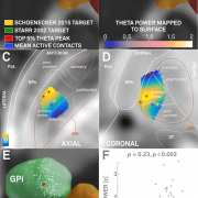
Inside a new version of Lead-DBS (v2.0.0.6) released yesterday, we included the pallidal theta activity sweet spot defined in A localized pallidal physiomarker in cervical dystonia as well as optimal stimulation targets defined in Thomas Schönecker‘s Article Postoperative MRI localisation of electrodes and clinical ef… and Philip Starr’s Article Microelectrode-guided implantation of deep brain stimulators….
We further included the CIT168 subcortical atlas defined in A High-Resolution Probabilistic In Vivo Atlas of Human Subco… that Wolfgang Pauli and colleagues from Caltech graciously shared for inclusion in Lead-DBS.


